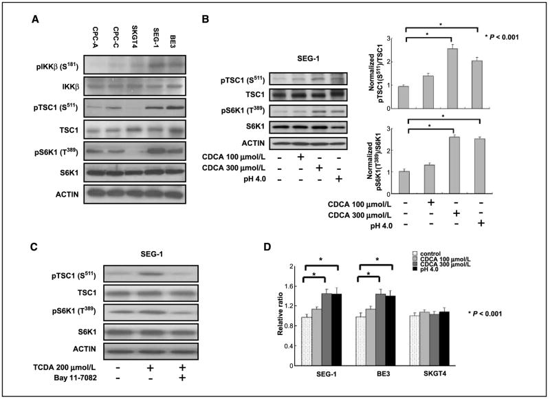
Figure 1.
IKKβ/TSC1/mTOR pathway was activated in Barrett’s esophagus–associated EAC cancer cells. A, increased expression of pIKKβ (S181) was correlated with higher level of pTSC1 (S511) and pS6K1 (T389) in SEG-1 and BE3 EAC cancer cell lines. Immortalized Barrett’s esophagus cell lines CPC-A and CPC-C had lower expression level of pIKKβ (S181) and expressed concurrently low levels of pTSC1 (S511) and pS6K1 (T389). The SKGT4 EAC cancer cell line also had lower expression of pIKKβ (S181), pTSC1 (S511), and pS6K1 (T389). B, the phosphorylation of pTSC1 (S511) and pS6K1 (T389) increased two to three times in SEG-1 cancer cells treated with 300 μmol/L CDCA (P < 0.001) and low-pH medium (P < 0.001) compared with those in untreated cells. C, conjugated bile acid TCDA increased the expressions of pTSC1 (S511) and pS6K1 (T389), and the IKKβ inhibitor Bay 11-7082 blocked the TCDA stimulation effect in SEG-1 EAC cancer cells. D, cell proliferation was increased in SEG-1 and BE3 cancer cells compared with SKGT-4 cells, which had lower expression of the IKKβ/TSC1/mTOR signaling pathway.

