Figure 1.
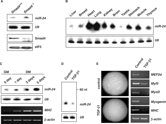
Suppressed expression of miR-24 by TGF-β1. (A) The expression of miR-24 in Smad4+/+ and Smad4–/– mouse heart tissues was detected by Northern blot. The expression of Smad4 was detected by Western blot. U6 RNAs detected by Northern blot and eIF5 were used as loading controls respectively. (B) Expression profile of miR-24 in 2-month old mouse tissues detected by Northern blot. (C) Northern blot analysis of miR-24 expression using total RNA isolated from C2C12 myoblasts cultured in growth medium (GM) or differentiation medium (DM) for 0, 1, 3 and 5 days. RT-PCR analysis of MHC expression to monitor the differentiation status at indicated times. (D) Northern blot analysis of miR-24 expression in C2C12 cells transferred to DM with or without TGF-β1 (5 ng/ml) for 3 days. (E) Morphological demonstration and differentiation marker expression (MEF2d, Myf5, MyoD, Myogenin, MHC) of C2C12 cells transferred to DM with or without TGF-β1 (5 ng/ml) for 3 days.

