Figure 5.
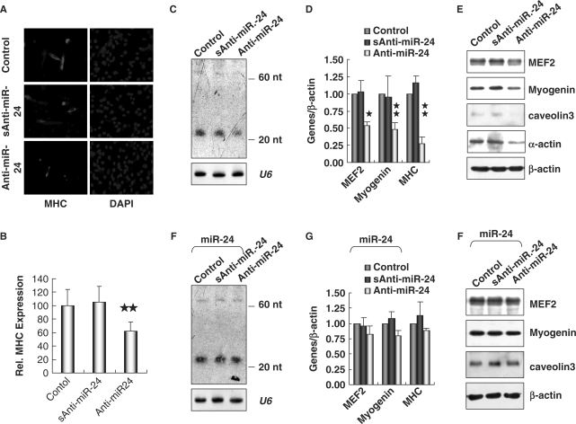
Inhibition of miR-24 suppressed myogenesis in C2C12 cells. (A) Knockdown of miR-24 inhibited the expression of MHC in myoblasts. C2C12 myoblasts were electroporated with 2′-O-methyl antisense inhibitor of miR-24 (Anti-miR-24) with scrambled sequence (sAnti-miR-24) as a negative control. The myoblasts were then cultured in GM for 24 h and transferred into DM for 36 h before immunofluorescence staining of MHC. DAPI staining was performed to visualize the nuclei. (B) The expression of MHC shown in (A) was quantified after standardization with the expression level of MHC in controls. Two stars mean P < 0.01. (C) The expression of miR-24 detected by Northern blot in C2C12 myoblasts electroporated with Anti-miR-24. U6 RNAs detected by Northern blot were used as loading controls. (D) and (E) Knockdown of miR-24 suppressed the expression of myogenic factors, which was assessed by real-time RT-PCR (D) and Western blot analyses (E). (F) C2C12 myoblasts were first infected with retroviral vector pINCO-miR24 and then electroporated with Anti-miR-24. The expression of miR-24 was detected by Northern blot. U6 RNAs detected by Northern blot were used as loading controls. (G) and (H) The expression of myogenic factors assessed by real-time RT-PCR (G) and Western blot analyses (H) in cells treated as (F). One star means P < 0.05, two stars mean P < 0.01.

