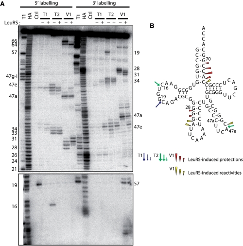
Figure 2.
Nuclease probing of AatRNALeuCAA in the free form or in complex with AaLeuRS. (A) tRNALeuCAA was labeled at its 5′-end or 3′-end. Probing was done in the presence (+) or absence (−) of LeuRS. OH and T1 are ladders of the tRNA under the denaturing condition; Ctrl is the control without any probe. The probes comprised RNase T1, RNase T2 and RNase V1. Numbers refer to tRNA nucleotide positions. (B) Cloverleaf structure of tRNA summarizing the reactivity changes observed in the tRNA following AaLeuRS binding. The symbols and color codes for the probes are indicated in the figure. Three intensities of cuts/modifications for each probe are shown (strong, medium and moderate).

