
Figure 1.
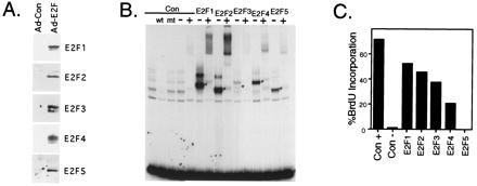
Induction of S phase by E2F family members. (A) Overexpression of each of the E2F family members in REF52 cells. REF52 cells were deprived of serum for 48 hr (0.25% serum) and then infected with either the control recombinant adenovirus (Ad-Con) or with a recombinant expressing the indicated E2F family member (E2F1, E2F2, E2F3, E2F4, or E2F5) at an moi of 75 ffu/ml for each virus. A virus expressing the DP1 protein, Ad-DP1, was coinfected (moi of 75 ffu/ml) with each E2F expressing virus. Following infection, the cells were returned to 0.25% serum media. The cells were harvested 15 hr postinfection for Western blot analysis using the corresponding antibody specific for the indicated E2F family member. (B) Production of E2F activity following infection with recombinant adenoviruses. Starved REF52 cells were infected with either the control recombinant adenovirus (Con) or with a recombinant expressing the indicated E2F family member at the following mois: Ad-Con, Ad-E2F1, Ad-E2F2, Ad-E2F4, and Ad-E2F5 at 400 ffu/ml, or Ad-E2F3 at 800 ffu/ml. Even at the elevated moi used for Ad-E2F3, less activity was produced than for the other E2Fs. Five hours postinfection, the cells were stimulated with 10% serum and then harvested 15 hr later for an electrophoretic mobility shift assay using a DHFR E2F binding site as a probe. Complexes specific for each overexpressed E2F are indicated with an asterisk. Where indicated, a polyclonal antibody against the DP1 protein (+) or control rabbit immunoglobulin (−) was added to the binding reaction after 20 min, and the reaction continued for an additional 20 min. Lanes 2 and 3 depict competition with either wildtype (wt) or mutant (mt) binding sites. (C) E2F family members induce S phase in quiescent cells. Serum starved REF52 cells were infected as described in Fig. 1B. Following infection, the cells were returned to either 10% serum media (Con+, left bar) or 0.25% serum media (other bars). Cells were labeled with BrdUrd from 12 to 30 hr, and BrdUrd incorporation was determined by indirect immunofluorescence. At least 200 nuclei were scored for BrdUrd incorporation.