
Figure 4.
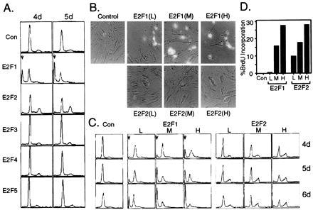
The unique capacity of E2F1 to induce apoptosis (A) E2F1, but not the other E2F family members, induces apoptosis. REF52 cells were deprived of serum for 48 hr, and then infected with the indicated recombinant viruses at an moi of 200 ffu/cell. Following infection, the cells were returned to 0.25% serum media and harvested at either 4 or 5 days postinfection for analysis by flow cytometry. The horizontal axis reflects relative DNA content, and the vertical axis represents cell number. The position of cells with less than a G1 DNA content, indicative of apoptosis, is shown by an arrow. (B) A comparison of the relative abilities of E2F1 and E2F2 to induce apoptosis. REF52 cells were deprived of serum for 48 hr and then infected with Ad-E2F1 or Ad-E2F2 at either a low (50 ffu/cell), medium (100 ffu/cell), or high (200 ffu/cell) moi. Alternatively, cells were infected with Ad-Con at 100 ffu/cell. Following infection, the cells were returned to 0.25% serum media and harvested at either 4, 5, or 6 days postinfection for analysis by flow cytometry. (C) E2F1, but not E2F2, expressing cells exhibit morphological characteristics of apoptosis. Cells from Fig. 4B were photographed at 5 days postinfection, and representative photographs are shown. (D) Both E2F1 and E2F2 induce S phase. Cells from Fig. 4B were also labeled with BrdUrd from 10 to 40 hr postinfection, and BrdUrd incorporation was determined as described in Fig. 1B.