
Figure 1.
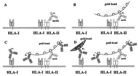
Schematic view of the steps of sequential labeling of the cell surface elements. (A) First specific labeling against antigens (HLA II) with mAb. (B) Larger gold beads attached to the Fc part of the antibody. (C) Second specific labeling of antigens (HLA I). These mAbs also cover the free binding sites of the gold beads. (D) Smaller size gold beads bind the Fc part of the second antibody.