
Figure 5.
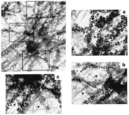
Cell whole mount electron micrograph of microtubules and protein 4.1 at a centrosome of a human fibroblast. Protein 4.1 epitopes (5-nm beads) and β-tubulin (10-nm beads) on microtubules were detected in fibroblasts prepared under conditions for microtubule stabilization. Three regions (a, b, and c), from the boxes on the image at low magnification, present examples of separate 4.1 epitope distribution (arrows) relative to microtubules. (Bar = 100 nm.)