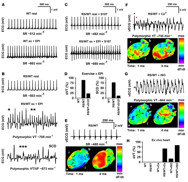
Figure 4. Fixing the leak in mutant channels from Ryr2RS/WT mice with S107 protects against fatal cardiac arrhythmias.
(A–C) Representative telemetric ECG recordings of WT (n = 6), heterozygous Ryr2RS/WT (n = 9), and Ryr2RS/WT mice treated with S107 (n = 9). (A) ECGs of a WT mouse, sedentary (rest) and after 45 minutes of treadmill running immediately followed by catecholamine injection (ex + EPI; epinephrine, 0.5 mg/kg i.p.). (B) ECGs of a Ryr2RS/WT mouse, sedentary and following arrhythmia provocation stress testing, which resulted in rapid sVT and SCD. *Bidirectional VT; **polymorphic VT; ***rapid polymorphic VT. (C) Ryr2RS/WT mice treated with S107 under sedentary housing conditions (rest) and following stress testing (S107, 5 mg/kg/h s.c. for 7 days by osmotic pump). S107 prevented stress-induced arrhythmias. (D) Occurrence of sVT (left) and SCD (right). (E) Example of Langendorff-perfused Ryr2RS/WT hearts (n = 6) that exhibited regular SR recorded by volumetric ECG (vECG) with 2 epicardial breakthroughs (arrows) and homogenous apical-to-basal voltage activation. (F) Epicardial voltage activation map of the same Ryr2RS/WT heart showing multiple activation foci (arrows) and abnormal activation wavefront propagation during rapid polymorphic VT. The black dot marks the last regular sinus beat of vECG, and the red dot the first abnormal beat occurring at a short coupling interval. (G) Ryr2RS/WT heart showing abnormal focal activation (arrows) rapidly moving toward apex and left ventricle during sVT. (H) Occurrence of sVT in ex vivo perfused WT and Ryr2RS/WT hearts (each n = 6). Perfusion in the absence or presence of either high extracellular Ca2+ (9 mM) or ISO (100 nM) resulted in sVT in 9 of 12 Ryr2RS/WT but only in 1 of 12 WT hearts. Orientation of the heart is as indicated; a, apex; b, base; l, left; r, right of anterior activation maps.

