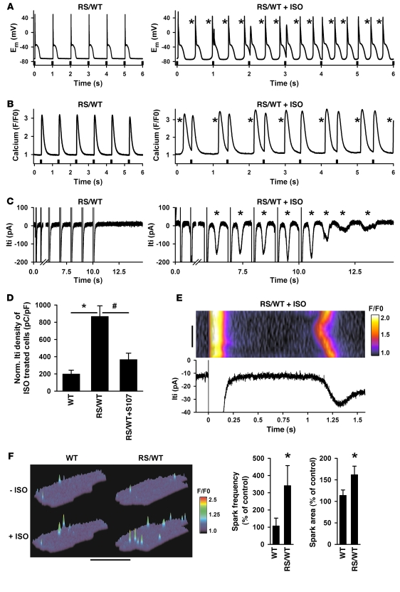
Figure 5. Electrical and Ca2+ cycling abnormalities in Ryr2RS/WT cardiomyocytes are consistent with Ca2+-triggered afterdepolarizations and are reduced by the RyR2-stabilizing drug S107.
(A) Representative examples of whole-cell current (Em) recordings from Ryr2RS/WT cardiomyocytes paced continuously at 1 Hz (recording started after 2 minutes; pacing stimuli as indicated) under control (left) and ISO-stimulated conditions (1 μM; right). Following ISO, aberrant membrane depolarizations (asterisks) increased in frequency and became self-sustained when pacing was stopped (not shown). (B) Intracellular Ca2+ transients from Ryr2RS/WT cardiomyocytes field-paced continuously at 1 Hz under control (left) and ISO-stimulated conditions (1 μM, right; recording started after 2 minutes continuous pacing). Aberrant spontaneous Ca2+ waves (asterisks) occurred irregularly and became self-sustained when pacing was stopped (not shown). (C) Representative current traces from Ryr2RS/WT cardiomyocytes recorded during an 0.5-Hz depolarization train in the absence (left) and presence of ISO (1 μM; right). ISO-treated Ryr2RS/WT cardiomyocytes showed frequent ITI (asterisks) during and after pacing. The figure illustrates currents recorded during and following the first 2 and last 5 pulses from a continuous 10-pulse (1 Hz) conditioning train. A 5.5-second-long period of the recording during the train was omitted for display purposes, and the time scale is expanded in the ISO-treated examples in the rights panels of A–C. (D) Normalized ITI density in ISO-treated cells. ISO-treated Ryr2RS/WT cardiomyocytes (n = 7) showed significantly increased ITI densities (*P < 0.05 vs. control; n = 6); in vivo S107 treatment significantly decreased ITI density in ISO-treated Ryr2RS/WT cells (#P < 0.05; n = 4). (E) Simultaneous confocal Ca2+ imaging of a small region of interest and ITI recording from an ISO-stimulated Ryr2RS/WT cardiomyocyte. Following regular pacing-induced Ca2+ release (last cycle shown on left side), intracellular Ca2+ and membrane current rapidly normalized to the resting (diastolic) state. Ryr2RS/WT cardiomyocytes showed abnormal intracellular Ca2+ release events, which became organized as Ca2+ waves, coinciding with secondary arrhythmogenic ITI. Scale bar: 10 μm; time and normalized F/F0 fluorescence signal are as indicated. (F) Representative confocal Ca2+ spark images from WT and Ryr2RS/WT cardiomyocytes before (–ISO) and after (+ISO; 1 μM) treatment. Bar graphs show significant differences (*P < 0.05) in average spark frequencies and average spatial area dimensions, indicating increased intracellular Ca2+ leak in Ryr2RS/WT cardiomyocytes following ISO stimulation as the subcellular origin of arrhythmogenic diastolic DADs (A), Ca2+ waves (B and E), and ITI (C and E) events.

