
Figure 3.
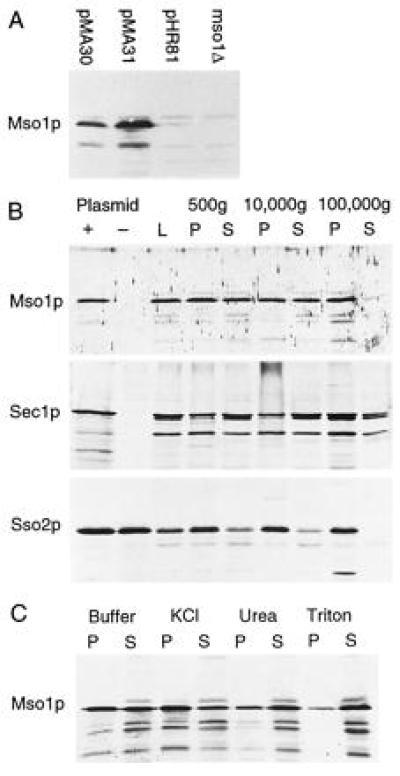
Western blots. (A) Detection of Mso1 protein in wild-type cells carrying either pMA30, pMA31, or the cloning vector pHR81. An mso1-disrupted strain is shown to the right. (B) Fractionation of yeast cell lysates by successive centrifugations at the indicated speeds. L, spheroblast lysate; P, pellet; S, supernatant. For Mso1p and Sec1p, whole cell lysates prepared in the presence and in the absence of the respective overexpression plasmid is shown (Left). There is no difference for Sso2p, because we did not need overexpression to detect this protein. (C) Membrane association of Mso1p in the 100,000 × g pellet. The pellet was treated either with Hepes buffer alone, or with buffer containing 1 M KCl, 2.5 M urea, or 1% Triton-X 100, respectively, and then separated by centrifugation at 100,000 × g.