
Figure 3.
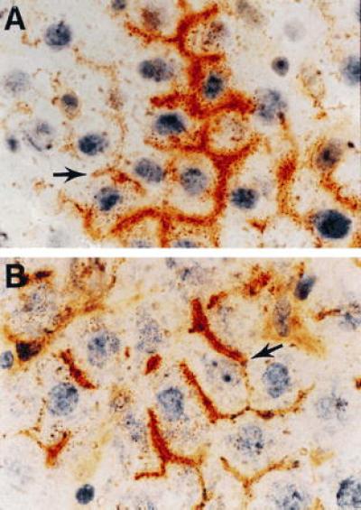
Identification of DPPIV-positive pancreatic and liver epithelial cells transplanted to the liver of DPPIV-negative mutant F344 rats. Dual histochemical staining of the tissue for DPPIV (orange/rust) and ATPase (brown) was conducted as noted. (A) Section of liver from a DPPIV-negative mutant F344 rat transplanted with an epithelial cell-enriched fraction from the pancreas of an 8-week-treated Cu-deficient DPPIV-positive F344 rat (×600). (B) Section of liver from a DPPIV-negative mutant rat transplanted with an epithelial cell-enriched fraction from the liver of a DPPIV-positive F344 rat, 2.5 days after treatment with d-galactosamine (×600). Arrows point to hybrid bile canaliculi formed between transplanted cells and endogenous hepatocytes.