
Figure 4.
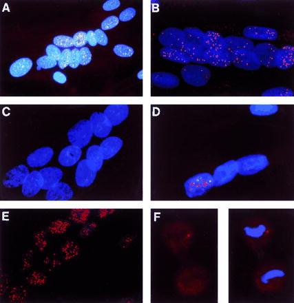
Distribution of triplet repeat transcripts in myoblasts and dividing cells. Fixed cells on coverslips were hybridized with cy3-labeled CAG-30 and counterstained with DAPI. Spots represent signal from the trinucleotide repeat. (A–E, ×225; F, ×300.) (A) Analog photomicrograph. (B–F) Digital images, generated by mathematically removing out-of-focus light from images captured by a CCD camera. Each digital image is a restored optical section through the cell(s). (A and B) DM myoblasts. (C) Normal myoblasts. (D) DM myoblasts from a minimally affected patient with approximately 150 CTG repeats. (E) DM myoblasts treated with DNase I and extracted with (NH4)2SO4. The expanded triplet repeat molecules remained in the nuclear matrix. The lack of DAPI staining indicates that the DNA has been removed. (F) Dividing fibroblasts from the same patient as in A. In late anaphase (possibly telophase), no signal was detected in the newly reformed nucleus, suggesting that transcript foci have diffused within the cytoplasm. (Left) Dividing cell without DAPI stain. (Right) DAPI shows the condensed DNA typical of anaphase.