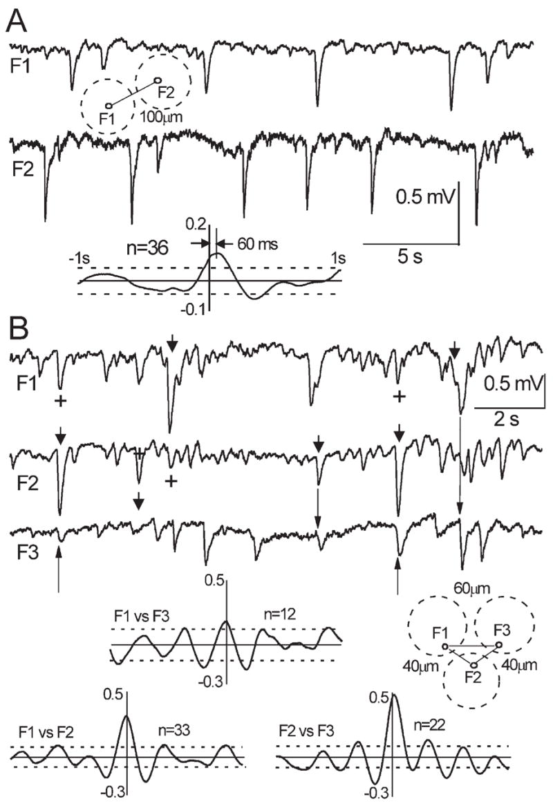
Fig. 5.

(A) Dual recording in a horizontal slice with an inter-electrode distance of 100 μm. Dashed circles indicate position of glomeruli. The averaged CCF (n = 36) shows significant correlation between sites with 60 ms time lag, indicating polysynaptically mediated propagation of excitation. (B) Triple recording in a horizontal slice with an inter-electrode distances: F1–F2 and F2–F3 40 μm, F1–F3 60 μm. Note utterly synchronous events, which are indicative of the field spread. Arrowheads designate bigger, presumably primary events; plus stands for presumably secondary events resulting from a field spread; arrows are pointed to slightly delayed sGLFP that could be initiated by primary sGLFP via interglomerular connections. The insets indicate positions of extracellular electrodes, dashed circles designate corresponding glomeruli; avgCCFs indicate passive propagation of the fields without time lags between the sites of recording. Note pronounced ~3 Hz patterns of background oscillations in F1 and F2 which also emerged in CCFs.
