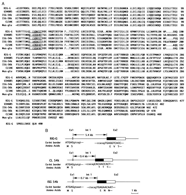
Figure 3.
Homology of RIG-G with related ISGs. (A) Comparison of deduced protein sequence of RIG-G with those of related ISG proteins. Amino acid residues conserved in the six ISGs are outlined. A potential tyrosine phosphorylation site is underlined. The numbers on the right represent the amino acid residues of the corresponding protein. (B) The exon-intron organization of RIG-G-related family members. The boxes represent the exons of the genes, and the coding regions of the genes are designated by filled boxes. The length of intron 1 is indicated by arrowheads. The exon-intron border sequences are listed below the exon-intron organization. The nucleotides of exon and intron are shown in capital and lowercase letters, respectively, and the corresponding amino acid residues are listed below the exon sequences.

