Fig. 3.
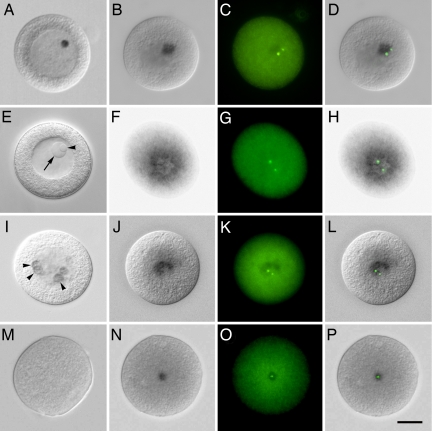
In situ localization of cnRNAs. (A) Differential interference contract microscopy (DIC) image of an unactivated oocyte labeled for cnRNA65. A dense circular patch within the GV is seen, similar in size and position to the nucleolinus. Note that both the nucleolinus and nucleolus are morphologically indistinct after cells were subjected to the hybridization regimen. A DIC image of a different unfixed oocyte is therefore shown in E for comparison with A. An arrow points to the nucleolus, and the nucleolinus is indicated by an arrowhead. B shows the distribution of cnRNA65 at 7 min postactivation. A distinct patch, although slightly more diffuse than seen in GV oocytes, is visible in the cytoplasm. Centrosomes (labeled with anti-γ-tubulin antibody in C) appear within or “attached to” the cnRNA hybridization patch. (D) Overlay of B and C. F (DIC) and G (immunofluorescence) show cnRNA65 and γ-tubulin, respectively, at 24 min postactivation (overlay in H). A series is shown for cnRNA15 in I–L: I, unactivated oocyte, arrowheads highlight ring-like hybridization patterns; J and K, cnRNA15 and γ-tubulin, respectively, at 7 min postactivation; L, overlay of J and K. (M) Hybridization control using cnRNA239 sense probe. (N) cnRNA239 (using antisense probe) at 7 min postactivation; O, γ-tubulin staining in the same cell; P, overlay. (Scale bar in P: 20 μm.]

