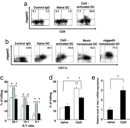
Fig. 5.
DC-mediated NK cell activation is controlled by Notch signaling. (a) The expression of Jagged2 on splenic DC from naïve or CpG-treated mice was evaluated by flow cytometry after staining cells with PE-conjugated anti-CD11c, FITC-conjugated anti-CD8α mAb, and biotin-conjugated anti-Jagged2 mAb, followed by streptavidin-APC. Data shown are gated on CD11c+ cells. (b) The expression of Jagged2 on bone marrow-derived untreated, CpG-treated, control vector-transduced, or Jagged2-transduced DC was evaluated by flow cytometry. Cells were stained with PE-conjugated anti-CD11c mAb and biotin-conjugated anti-Jagged2 mAb, followed by streptavidin-APC. (c) NK cells from BALB/c mice were cultured with Jag2-DC in the presence (black bars) or absence (gray bars) of 10 μM GSI. The cytotoxic ability of NK cells against YAC-1 cells was measured by 51Cr release from YAC-1 cells. The activity of NK cells without Jag2-DC stimulation was used as a control (white bars). Data are shown as the mean ± SD. *, statistical significance (P < 0.05). (d) NK cells from BALB/c mice were cultured with unstimulated or CpG (2 μM)-stimulated bone marrow-derived DC for 16 h in the absence (white bars) or presence (black bars) of 10 μM GSI. NK cells were enriched and cocultured with 51Cr-labeled YAC-1 cells, and 51Cr release from YAC-1 cells was measured. Data are shown as the mean ± SD. *, statistical significance (P < 0.05). (e) NK cells from BALB/c mice were cultured with unstimulated or CpG (2 μM)-stimulated bone marrow-derived DC for 16 h. The mRNA expression of HES1 in purified NK cells was quantified by real-time PCR. The value obtained from Notch1 expression in NK cells cocultured with unstimulated DC was set as 1. Data are shown as the mean ± SD. *, statistical significance (P < 0.05).

