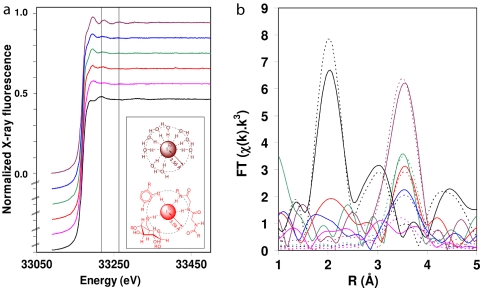
Fig. 1.
XAS of L. digitata tissues, including time course of experiment with fresh Laminaria thalli stressed with oligoguluronates (GG). (a) Iodine XAS (K-edge region). (b) Phase-corrected FT of k3-weighted EXAFS (cf. Fig. S1). Solid lines, experimental; dashed lines, simulations. (Inset) Schematic representation of a cross-section of the immediate environment of the I− ion, showing the change in the solvation by many hydrogen-bonded H2O molecules in 20 mM NaI solution (Upper) to association by fewer hydrogen bonds to biomolecules in fresh Laminaria (Lower; examples clockwise from top left are polyphenol, peptide, and carbohydrate). In Laminaria tissues, iodine is overwhelmingly stored as iodide. In fresh, live tissues, iodide is largely present in association with biomolecules including polyols (e.g., carbohydrates), phenols (e.g., phlorotannins), and amides (e.g., proteins), replacing the highly ordered hydration shell. Upon oxidative stress, part of this iodide is mobilized, as reflected in a more ordered hydration shell. When freeze-dried tissues are exposed to 2 mM hydrogen peroxide in seawater (simulating local concentrations observed during an oxidative burst), much of the available iodine is incorporated into aromatic organic molecules as reflected by the strong change in the EXAFS spectrum (highlighted by the vertical lines at 33,219 eV and 33,266 eV, respectively). Black, lyophilized Laminaria rehydrated in seawater with 2 mM H2O2 [simulation: 1.0 phenyl at 2.1 Å, a = 0.003 Å2 (a, Debye–Waller-type factor as 2σ2 in Å2)]; pink, lyophilized Laminaria (2.2 O at 3.51 Å, a = 0.038 Å2); red, fresh Laminaria (1.6 O at 3.58 Å, a = 0.005 Å2); sea green, 20 min after exposure to GG (2.8 O at 3.58 Å, a = 0.021 Å2); blue, 3 h after exposure to GG (3.9 O at 3.56 Å, a = 0.038 Å2); plum, 20 mM NaI (10.0 O at 3.56 Å, a = 0.034 Å2). A complete list of all simulation parameters with fitting errors is available as Table S1.

