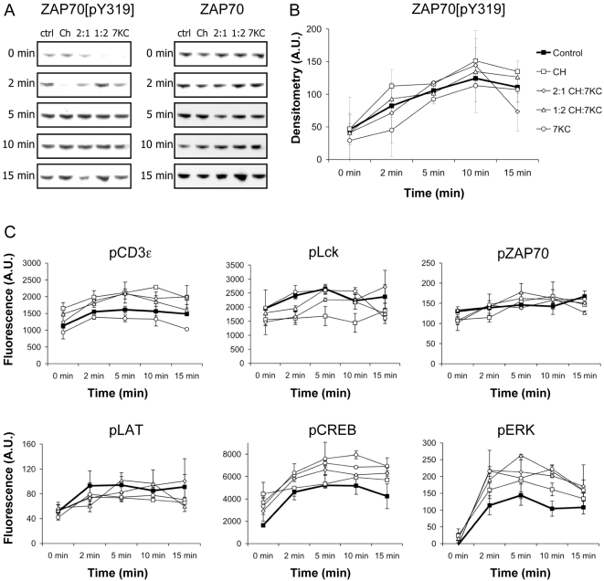
Figure 5. Signaling activities upon TCR triggering in whole cell lysates.
A. Control and sterol-enriched Jurkat cells were activated with 5 μg of UCHT1 (anti-CD3 mAb) for the indicated periods of time. Whole cell lysates were probed for ZAP70 phosphorylated at tyrosine 319. B. Quantification of tyrosine 319 phosphorylation of ZAP70. The data show the mean and range of two independent experiments. C. Multiplex analysis of T cell signaling. 2×10⁶ sterol-enriched Jurkat wt cells were activated with 5 μg/ml of anti-CD3 UCHT1 antibody for 0-15 min at 37°C. Non-site specific tyrosine phosphorylation of CD3ε, Lck, ZAP70, LAT and ERK1/2 (Tyr185/Tyr187) as well as serine phosphorylation of CREB (Ser133) in whole cell lysates was assessed by multiplex microbead suspension assay. The data is one representative experiment; error bars represent standard deviations. Legend shown in B applies to data in C.

