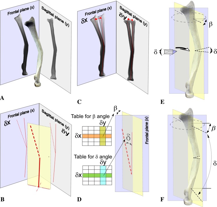
Fig. 3A–F.
A radius malunion consists of an angulation at the middle third of the bone in the radial-dorsal to ulnar-volar plane. The orientation of the deformity in space and the value of the maximal angular deformity, termed true angle of deformity, are assessed with (A) orthogonal radiographs. (B) The projections of the deformity in frontal and sagittal planes are shown as assessed with an orthogonal radiograph. (C) Preoperative planning is started with superposition of the radiograph of both sides. This allows assessment of the angular deformity in frontal (δx) and sagittal planes (δy). δx and δy are used to assess the value of the true angle of deformity (δ) and the orientation of the deformity in space (β) using the established table (D) [29]. (E) δ also defines the angle of the bone wedge that must be removed for a closed wedge osteotomy or the wedge of the structural bone graft to be inserted in an open wedge osteotomy to correct the deformity. (F) The correction must be performed in the plane of maximum deformity, defined by β in respect to the frontal plane. Intraoperatively, two Kirschner wires (plain line) are placed in the frontal plane using the distal radius as a landmark. The level of the osteotomy also is marked with a Kirschner wire. Subsequently, the plane of correction is marked with two Kirschner wires (dotted line) inserted with a β angle in respect to the Kirschner wires in the frontal plane. The second of these wires is inserted with a δ angle in respect to the first one. After completion of the osteotomy, the two Kirschner wires must be parallel (δ = 0) but the β angle is still the same.

