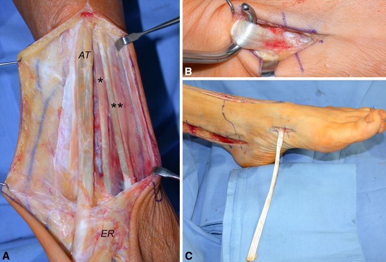
Fig. 2A–C.
These intraoperative photographs highlight the proposed procedure. (A) The tibialis anterior tendon (ATT; AT), extensor hallucis longus tendon (*), and extensor digitorum longus (**) tendons are exposed from muscular junction to the extensor retinaculum (ER). (B) The ATT bony insertion is identified. (C) The ATT is extracted distally.

