Fig. 1.

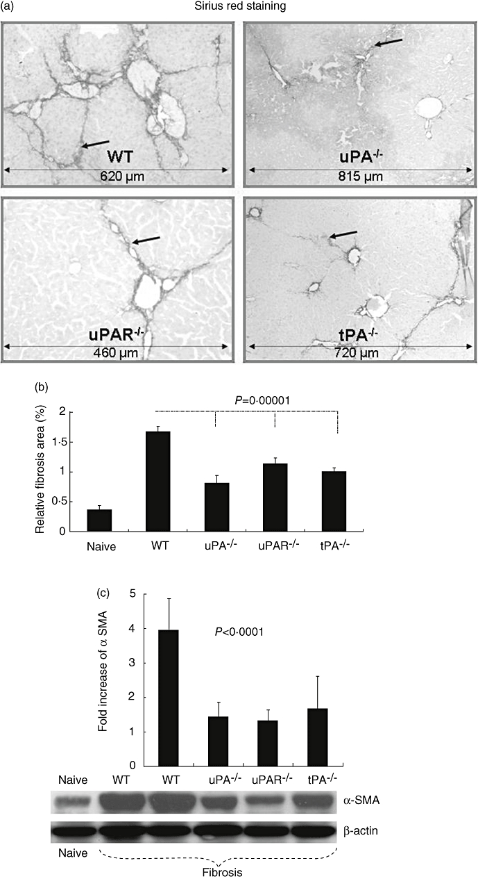
Reduced fibrosis in urokinase type plasminogen activator knockout (uPA−/−), uPA receptor (uPAR−/−) and tissue PA (uPA−/−)versus wild-type (WT) animals in a carbon tetrachloride model of liver injury. Tissue sections were stained with Sirius Red, as described in Materials and methods. Representative tissue sections are shown (a). Fibrotic septa, which are more established in WT than in −/− animals, are highlighted by arrows (scale bars are illustrated in the bottom of each field and presented with μm). Relative fibrosis area, expressed as percentage of total liver area, was assessed by analysing 36 Sirius Red-stained liver sections per animal (b). Each field was acquired at 10× magnification and then analysed using a computerized Bioquant® morphometry system. The relative fibrosis area in the livers of −/− animals was lower than that seen in WT mice. P-values refer to comparisons between WT and –/– animals after carbon tetrachloride. Whole liver protein lysates were extracted and 30 μg total protein was loaded per lane and analysed for alpha smooth muscle actin (αSMA) and β-actin expression (c). Decreased αSMA expression (lower panel) was found in lysates from uPA−/−, uPAR−/− and tPA−/− compared with WT receiving carbon tetrachloride and compared with naive WT animals. To obtain standardizations of β-actin/αSMA expression of all tested wells, bands were scanned and quantified as described in Methods. Results were expressed as fold increase compared with naive mice from each gel and (c, upper panel). The findings are representative of at least four different experiments with the same number of eight to 10 animals in each subgroup.
