
Fig. 5.
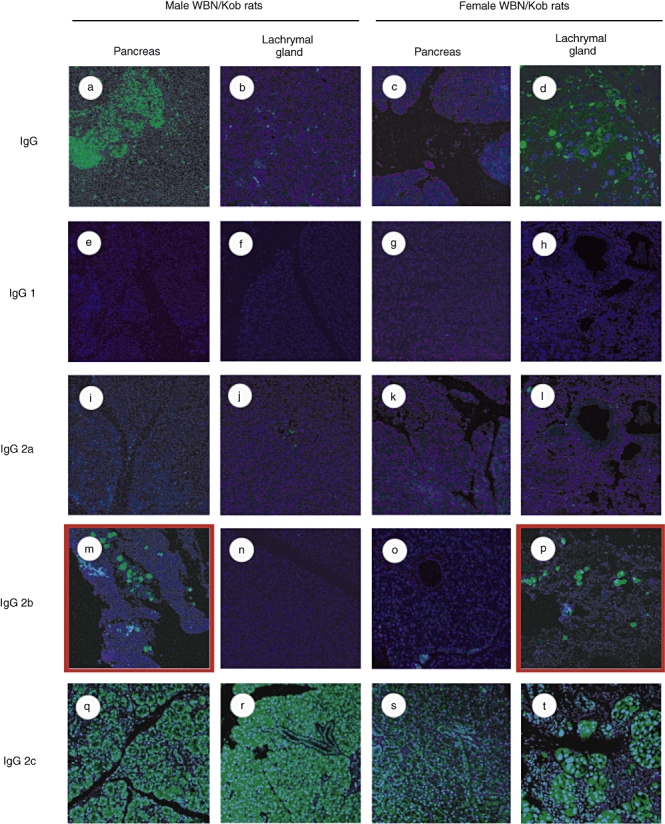
Detection of tissue-specific antibody. Paraffin sections of pancreas and lachrymal gland were prepared from male and female Wistar Bonn/Kobori (WBN/Kob) rats (4 months of age) and stained with biotinylated anti-IgG (a–d), anti-IgG1 (e–h), anti-IgG2a (i–l), anti-IgG2b (m–p) or IgG2c (q–t). They were visualized by additional staining with fluorescein isothiocyanate–streptavidin.