
Figure 3. Single Particle Helical Reconstruction of a CIP4 F-BAR Domain-Induced Membrane Tubule.
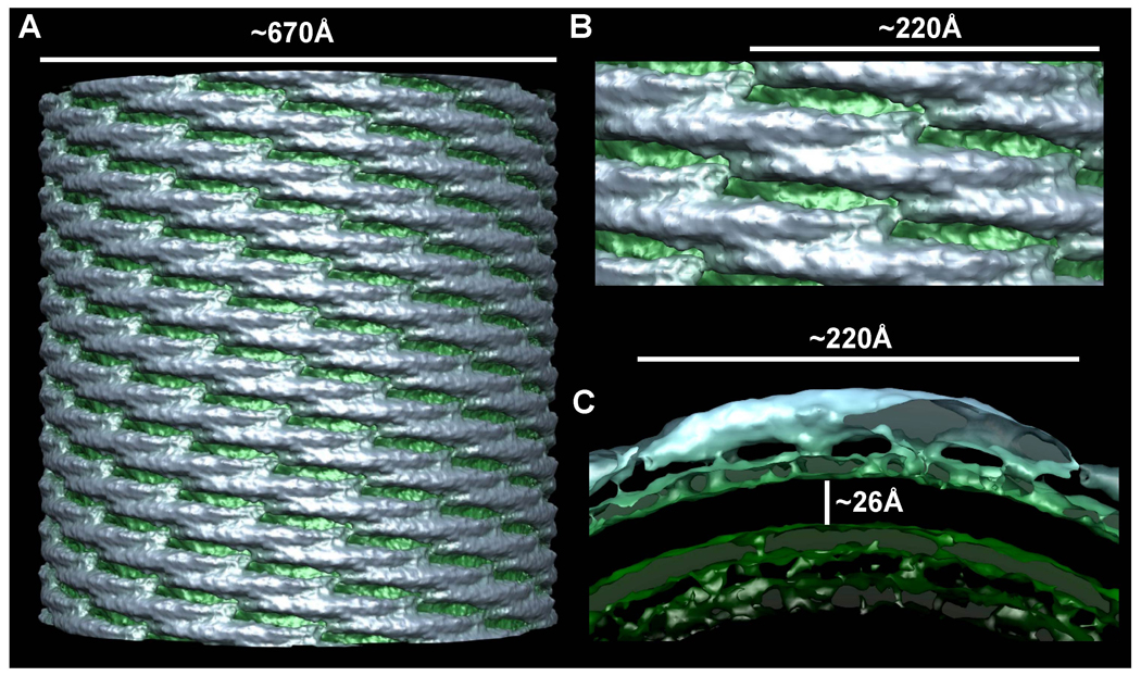
A) Surface of a ~67 nm diameter membrane tubule at ~17 Å resolution. The protein coat is colored blue-gray and the underlying membrane is green. B) Zoom in on the lattice seen orthogonal to the cylindrical axis, highlighting the tip-to-tip interactions and the broad contacts between laterally-adjacent dimers. C) Cross-sectional slab through one dimer parallel with the plane of the tip-to-tip interaction. There are four clearly resolved points of membrane binding. The hydrophobic core of the phospholipid bilayer is ~26 Å thick and the headgroup regions are ~12Å thick.