
Figure 5. Independent Reconstructions of Tubules with Different Diameters and Symmetries.
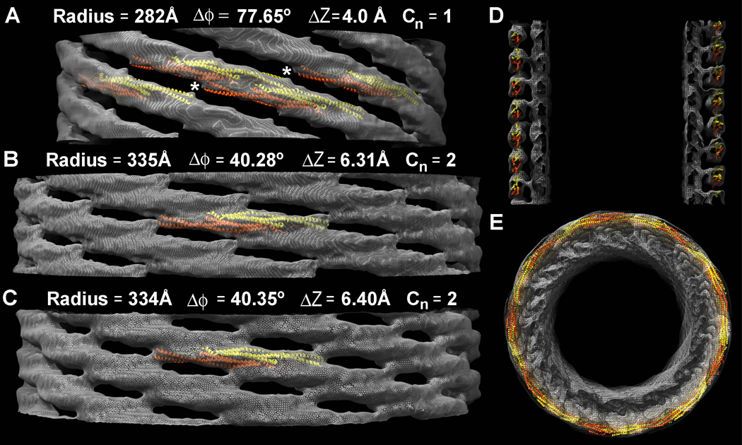
A) The narrowest tubule reconstructed is ~56 nm in diameter, with ~8 tip-to-tip dimers around its circumference. Tilting the long axis of the dimer relative to the cylindrical axis produces a narrower tubule. In this case, the dimers are so steeply tilted that the tip-to-tip contacts appear to be broken (white asterisks). The tubule has no rotational symmetry; the fundamental (J+1) helical symmetry does not describe an inter-molecular contact. Only the near side of the lattice is shown and the underlying membrane has been masked out to emphasize differences in the protein coat. Atomic models of F-BAR domains were fit into the map as rigid bodies. B&C) Two tubules with the same apparent diameter and ~9.5 tip-to-tip dimers around their circumference have resolvable differences in their helical symmetry. D) Central section along the longitudinal axis of the thinnest tubule shown in ‘A’, demonstrating that the density of the protein coat accommodates rigid atomic models of the F-BAR module that are tilted relative to the cylindrical axis, but whose radius of curvature is unchanged. E) View along the cylindrical axis of the thinner reconstruction shown in ‘A’ and ‘D’.