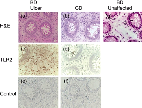
Fig. 2.

Characterization of Toll-like receptor (TLR)-2-expressing cells in the affected site of intestinal Behçet's disease (BD). (a) Haematoxylin and eosin (H&E) staining of an intestinal lesion of BD. (b) H&E staining of an intestinal lesion of Crohn's disease (CD). (c) Immunohistochemical staining with anti-TLR-2 monoclonal antibodies (mAb) of the affected site of intestinal BD. (d) Immunohistochemical staining with anti-TLR-2 mAb of the affected site of CD. We found hardly any TLR-2-positive cells in the lesion in this CD patient. (e) Immunohistochemical staining with control mouse immunoglobulin G (IgG) of the affected site of intestinal BD. (f) Immunohistochemical staining with control mouse IgG of the affected site of CD. (g) H&E staining of the unaffected site of resected intestine in patients with intestinal BD, where infiltrating mononuclear cells were detected marginally.
