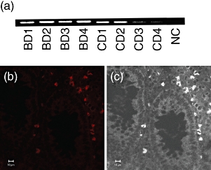
Fig. 5.

Heat shock protein (HSP) expression of intestinal lesions of Behçet's disease (BD). (a) Reverse transcription–polymerase chain reaction (RT–PCR) analysis of HSP60 expression of the intestinal lesions of BD patients. Intestinal specimens were used to extract total RNA, and the RNA was reverse-transcribed for PCR amplification. Intestinal lesions of four BD patients and four Crohn's disease (CD) patients were analysed. Beta-actin PCR was the same as Fig. 1d, and was thus omitted. NC, negative control. (b) Confocal microscopic analysis of HSP60 expression of the intestinal lesions of BD patients. (c) White/black image of (b) shows the location of HSP-expressing cells. Scale bars represent 10 μm for (b) and (c).
