
Fig. 3.
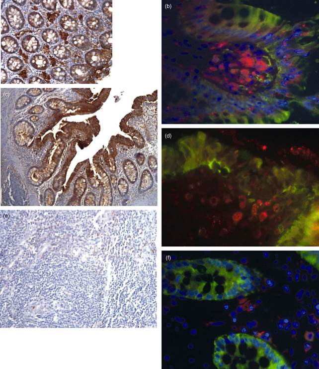
Immunofluorescence and immunohistochemical staining of galectin-3 (gal-3) in inflammatory bowel disease (IBD) colonic tissue, macrophages and controls. Three to five patients per group were investitgated. Representative staining of each group is shown in this figure. Gal-3 staining brown, CD 68 (macrophages) red, gal-3 and CD 68 orange. (a) Gal-3 staining in colon tissue of controls (brown, 200×). Gal-3 is expressed homogenously in the epithelial cell layer. (b) Gal-3 expression in macrophages (fluorescein isothiocyanate-tr-dapi, CD 68 red). (c) Gal-3 in ulcerative colitis (UC) colon (100×). (d) Gal-3 was found equally in UC macrophages. (e) Gal-3 expression in fistulae of Crohn's disease (CD) (200×). No gal-3 staining was found in this area. (f) Gal-3 in CD macrophages. Compared with control and UC patients there was no gal-3 expression in CD fistulae and a reduced expression in CD macrophages.