
Fig. 2.
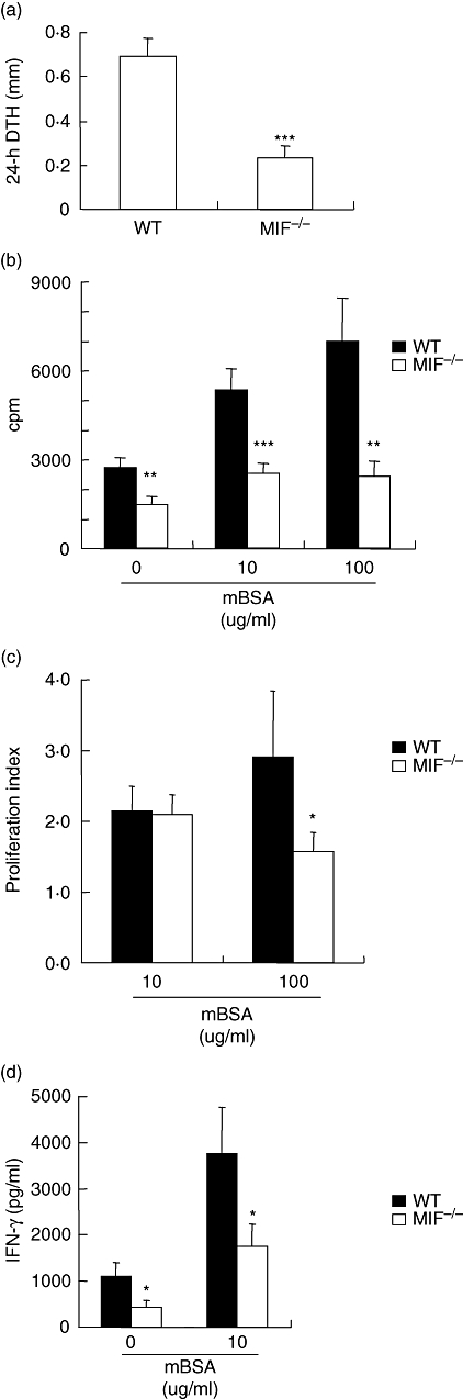
T cell activation in migration inhibitory factor (MIF) −/− and wildtype (WT) mice. Cutaneous delayed type typersensitivity was induced in sensitized mice by intradermal challenge with methylated bovine serum albumin (mBSA) in the footpad and footpad swelling measured 24 h later. Results are expressed as the difference between mBSA and saline-injected footpad thickness measured using micro callipers (a). Splenocyte proliferation was measured by [3H]-thymidine incorporation after 48 h in the presence or absence of mBSA (10, 100 μg/ml). Results are expressed as cpm (b) and proliferation index (see methods) (c). In a separate experiment, IFN-γ (d) from splenocyte culture supernatants was measured by enzyme-linked immunosorbent assay. MIF −/− mice had significantly decreased evidence of T cell activation in all parameters measured. All results are expressed as mean ± standard error of the mean of at least 7 mice in each group (*P < 0·05, **P < 0·01, ***P < 0·001). cpm, counts per minute; IFN, interferon.