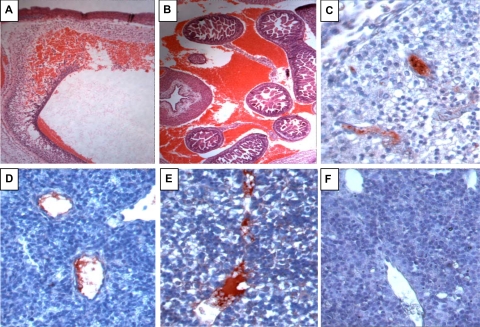
Figure 3.
Hemorrhage and intravascular coagulation in E13.5 FV(λ/λ)/ZPI(−/−) embryos. A-E, FV(λ/λ)/ZPI(−/−) embryos; F, FV(λ/λ)/ZPI(+/+) embryo. A,B, hematoxylin-eosin staining; C-F, antifibrinogen/fibrin staining (red) developed using AEC with hematoxylin counterstaining. (A) Intracranial hemorrhage (×5). (B) Intra-abdominal hemorrhage (×5). (C-E) Intravascular fibrin in the vessels of the brain (C, ×40), lung (D, ×40), and liver (E, ×40). In addition to the intravascular thrombosis, note disturbed cellular architecture and parenchymal fibrinogen/fibrin staining in the liver of the FV(λ/λ)/ZPI(−/−) embryo (E) compared with the liver of the FV(λ/λ)/ZPI(+/+) embryo (F).

