
Fig. 2.
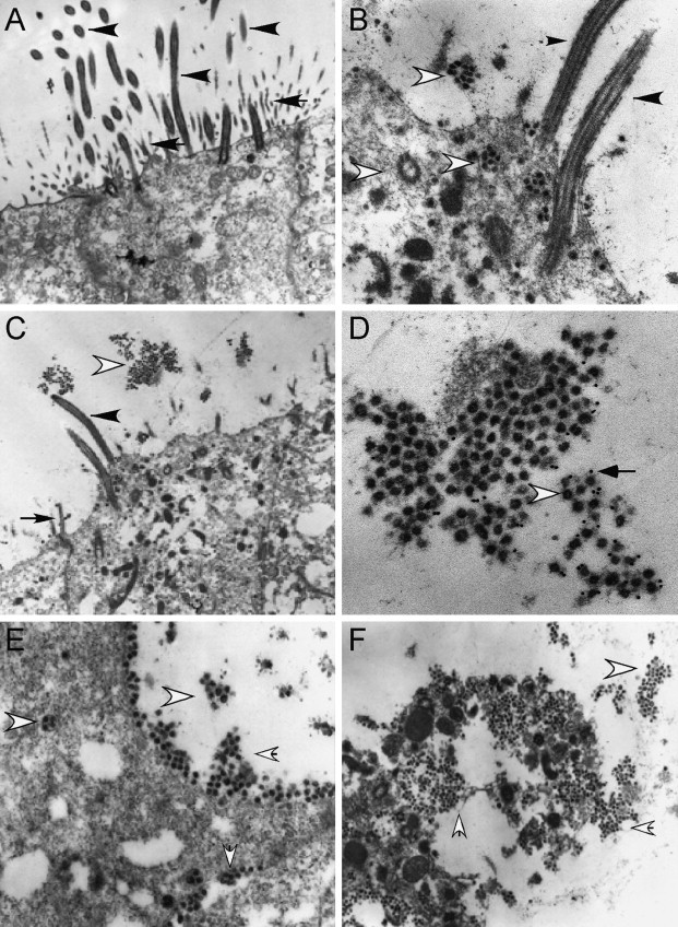
Ultrastructural localization of SARS-CoV in HAE. Representative transmission electron microscopic photomicrographs of HAE infected with Urbani SARS-CoV. (A) HAE inoculated with vehicle alone demonstrating the typical morphological features of the apical surfaces of ciliated cells with prominent cilia (black arrow heads) and microvilli (arrows). (B–F) HAE inoculated with Urbani SARS-CoV 48 h before fixation and showing the presence of large numbers of virus particles (open arrow heads) in vesicles inside the ciliated cells (B and E), and on the surface of ciliated cells (B–F). To confirm the observed virions were SARS-CoV, immuno-EM was performed using polyclonal mouse antisera against S with secondary antibodies conjugated to 12 nm gold beads (D, open arrow heads indicate virions, arrows indicate colloidal gold). SARS-CoV infection resulted in extrusion and shedding of infected ciliated cells into the airway surface microenvironment (F). Similar observations were seen with HAE infected with icSARS-CoV and SARS-CoV GFP. Black arrowheads, cilia; black arrows, microvilli; open arrowhead, virions; small arrow, immuno-EM colloidal gold.