
Fig. 3.
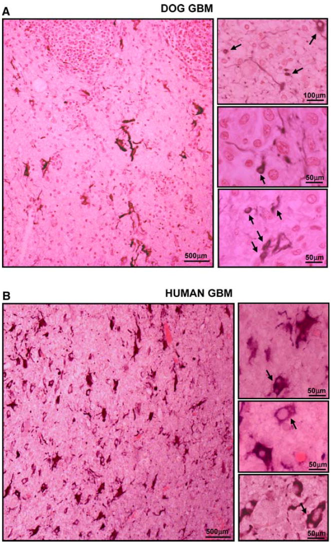
Neoplastic cellular infiltration into surrounding non-neoplastic brain tissue in spontaneous canine (a) and human GBMs (b). Paraffin sections (5 μm) from dog and human GBMs were stained with with GFAP antibodies. The microphotographs show that several of the small rounded cells infiltrating the non-neoplastic brain parenchyma express GFAP