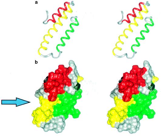
Figure 3.
(a) A stereo view of the backbone of RAPd1T structure. Helix H1 is red, H2 is yellow, and H3 is green. The unordered parts (see legend to Fig. 2) of the molecule are shown as thin gray lines. (b) The water-accessible surface of the RAPd1T structure. The color code is as in a. The arrow points to the position of the groove that may be the structural component involved in receptor binding. The figure has been prepared using insight ii (Biosym Technologies, San Diego).

