
Figure 9.
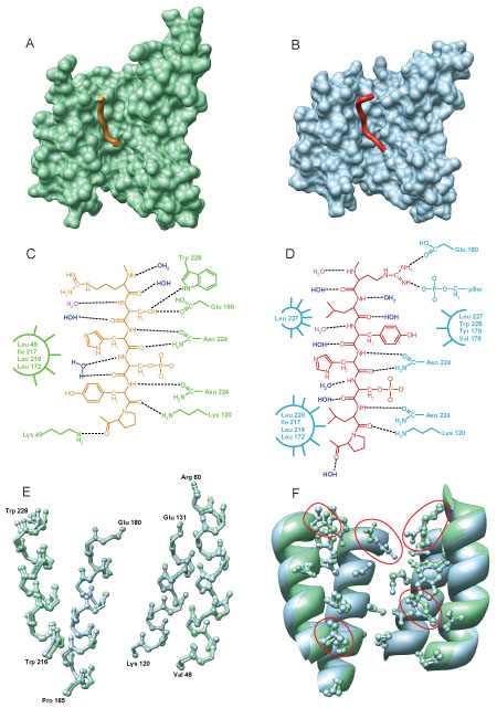
Detailed analysis of 14-3-3ζ peptide binding. The m1 peptide (A, orange ribbon) and m2 peptide (B, red ribbon) bound to 14-3-3 (A and B, shown by the green and blue surface, respectively). Details of 14-3-3 peptide binding are shown by a chemical schematic for the m1 peptide (C) and the m2 peptide (D), where both crystallographic waters (blue) and implicit waters (red) are shown. (E) Superposition of the backbone atoms from the 4 helices with the primary peptide binding residues for m1 (green) and m2 (blue) bound 14-3-3. (F) Superposition of ribbons of the same 4 helices showing the side chains of the residues that participate in m1 (green) and/or m2 (blue) binding.