Figure 1.
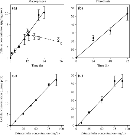
(a and b) Kinetics of uptake of telavancin (filled symbols and continuous line) in J774 macrophages (a) or embryo fibroblasts (b) incubated for the indicated times with an extracellular concentration of 90 mg/L at 37°C in medium supplemented with 10% fetal calf serum. (a) Kinetics of efflux of the drug from J774 macrophages exposed to telavancin (90 mg/L) for 12 h and re-incubated in a drug-free medium for an additional 24 h (open symbols and broken line) are also shown. (c and d) Cellular concentration of telavancin in J774 macrophages (c) or embryo fibroblasts (d) incubated at 37°C for 24 h or 72 h, respectively, in the presence of telavancin at the extracellular concentrations indicated on the abscissa. Results are given as arithmetic means ± SD (n = 3) and analysed by linear regression to calculate the corresponding clearances (µL/mg of protein/h): J774 macrophages, 9.6 ± 0.6 (R2 = 0.98; a) and 10.0 ± 0.4 (R2 = 0.99; c); fibroblasts, 8.2 ± 0.4 (R2 = 0.97; b) and 9.0 ± 0.7 (R2 = 0.98; d).

