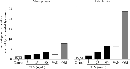
Figure 4.
Morphometric analysis of the material accumulated in macrophages (left-hand panel) or fibroblasts (right-hand panel) after 24 and 72 h of incubation, respectively, in control conditions or in the presence of 5, 25 or 90 mg/L telavancin (TLV), 50 mg/L vancomycin (VAN) or 20 mg/L oritavancin (ORI). Results are expressed as percentage of the cell surface occupied by the electron-dense material and/or large vesicles filled with a material of undetermined nature. Surface analysed was ~2000 µm2 for macrophages and 1000 µm2 for fibroblasts.

