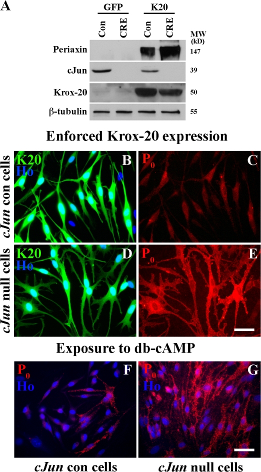
Figure 2.
Genetic removal of c-Jun amplifies Krox-20 or cAMP-induced myelin protein expression. (A) Western blot showing that c-Jun is absent from Junfl/fl cells infected with CRE-expressing adenovirus. The blot also compares periaxin in control (Con) and c-Jun–null cells (CRE) infected with GFP control adenovirus (GFP) or a Krox-20/GFP virus (K20). Note high periaxin levels in Krox-20–infected c-Jun–null cells (CRE). (B–E) c-Jun control (cJun con) and c-Jun–null mouse Schwann cells 2 d after infection with Krox-20/GFP adenovirus. Note that Krox-20 induces much higher levels of P0 protein in c-Jun–null cells (D and E) than in control cells (B and C). The reason why P0 levels in the Krox-20–expressing control cells appear low in this picture (C) compared with other comparable experiments (e.g., Fig. 4 I) is that exposure had to be reduced (equally for C and E) to avoid overexposure in E. (F and G) P0 protein expression in control cells P0 (cJun con) and c-Jun–null mouse Schwann cells after 3 d of exposure to db-cAMP/NRG-1. Note that cAMP/NRG-1 induces substantially higher P0 levels in cells without c-Jun. Bars, 15 μm.

