Figure 7.
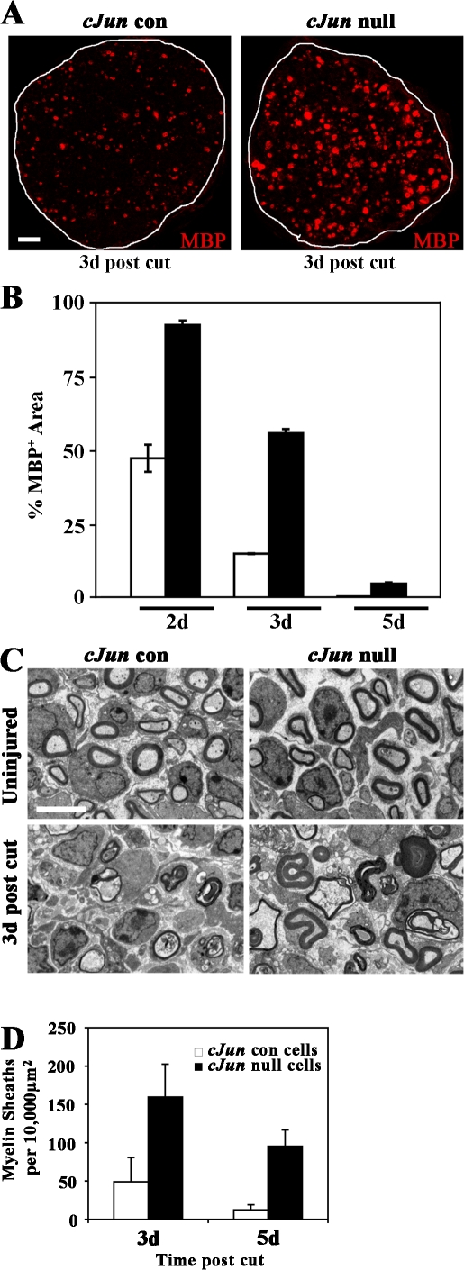
c-Jun drives dedifferentiation in vivo. (A) MBP immunolabeling of sciatic nerve sections showing delayed loss of myelin in c-Jun–null nerves compared with controls, 3 d after transection of nerves of 5-d-old mice. Bar, 10 μm. (B) Quantification of the delay in myelin disappearance by quantitative image analysis of MBP-immunolabeled sections (comparable to those shown in A) 2, 3, and 5 d after injury (expressed as percentage of MPB+ area in uncut P5 nerve). In every case, the difference between c-Jun–null and control nerves is significant (P < 0.01). (C) Electron micrographs showing c-Jun–null and control nerves from 5-d-old mice, intact and 3 d after injury as indicated. Note preservation of rounded or partially collapsed myelin sheaths in c-Jun–null nerves. Bar, 4 μm. (D) Counts of myelin sheaths (rounded or collapsed) in c-Jun–null and control nerves 3 and 5 d after injury (3 d, P < 0.05; 5 d, P < 0.01). Error bars show standard deviation of the mean.

