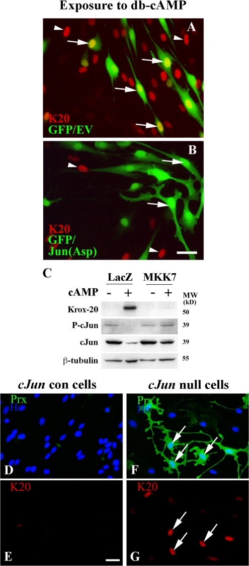
Figure 8.
Cross-inhibitory relationship between c-Jun and Krox-20. (A and B) Cells cotransfected with empty GFP vector (to visualize transfected cells) and an empty control vector (EV; A) or a Jun(Asp) vector (B). Both cultures were then treated with 1 mM db-cAMP for 2 d to induce Krox-20 and were immunolabeled for Krox-20. In A, arrows point to induced Krox-20 in nuclei of GFP-positive control cells (yellow nuclei of Krox-20–positive GFP-positive cells). In B, no Krox-20 is induced (arrows) in cells containing Jun(Asp). Arrowheads in both panels indicate untransfected cells that have been induced to express Krox-20 by db-cAMP as controls for induction. (C) Activation of JNK inhibits induction of Krox-20. Western blot of cells infected with adenovirus expressing control LacZ or virus expressing activated MKK7 to activate JNK is shown. Note that the Krox-20 and periaxin induced by 2 d of exposure to 1 mM db-cAMP in LacZ control cells is inhibited by MKK7 expression. Note also that MKK7 elevates c-Jun in the presence of db-cAMP. (D–G) In c-Jun–null cells, loss of Krox-20 expression is significantly delayed. Double immunolabeling of c-Jun control cells (D and E) and c-Jun–null cells (F and G) for Krox-20 (red) and periaxin (green) after 2 d in culture in DM containing 20 ng/ml NRG-1 is shown. Note that Krox-20 has disappeared from the control cells, whereas many c-Jun–null cells still have Krox-20–positive nuclei (G, arrows). Note that c-Jun–null Krox-20–positive cells are also periaxin positive (F, arrows), whereas control cells have lost periaxin expression (D). Bars, 15 μm.

