Figure 2.
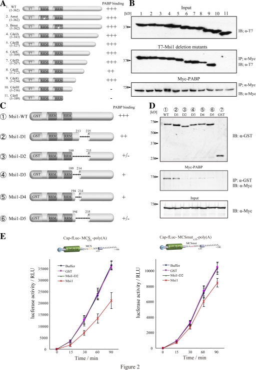
The C-terminal region of Msi1 that bound PABP is necessary for its function. (A) Illustration of proteins containing the T7-Msi1 variants: Msi1Amut (mutation in RRM1, and fails to bind mRNA: lane 2), Msi1Bmut (mutation in RRM2: lane 3), and a series of Msi1C-terminal deletions (lanes 4–11). (B) Immunoprecipitation using the T7-Msi1 variants was performed and various T7-Msi1 mutants bound to Myc-PABP (middle). The intensities of binding with PABP are illustrated to the right of panel A. (C) Illustration of the GST-Msi1 variants. (D) GST-Msi1 variants or GST as a control were coimmunoprecipitated with Myc-PABP in 293T cells using glutathione-Sepharose 4B (middle). PABP bound to Msi1 variants was immunoblotted using an anti-Myc antibody and is indicated (middle). (E) The in vitro–transcribed reporter mRNAs are illustrated at top (left, mRNA containing MCS; right, mRNA containing MCSmut), were translated in RRL with equimolar amounts of purified various GST proteins, and the luciferase activity was measured at each time point (0–90 min). The values represent mean ± SD; n = 5.

