Figure 4.
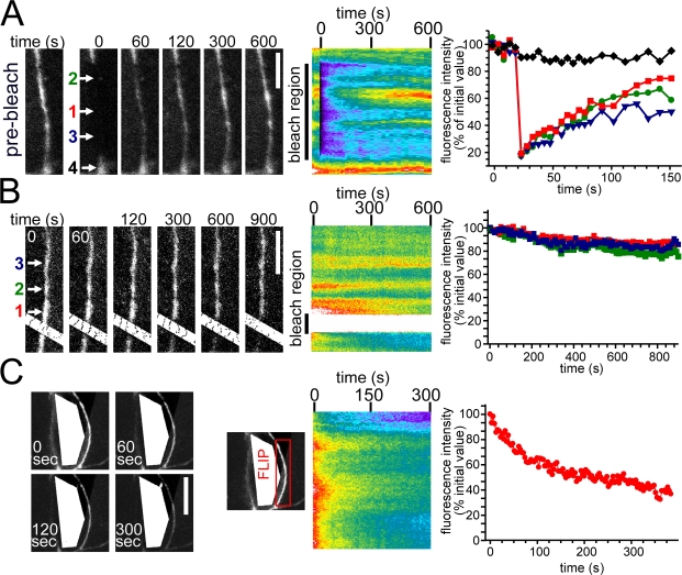
Tight junction–associated ZO-1 exchanges with an intracellular pool. (A) EGFP–ZO-1–expressing cells within confluent monolayers were studied by FRAP after photobleaching elongated tight junction regions. Representative images before and at the indicated times after photobleaching and the corresponding kymograph are shown. (B) The effect of continuous photobleaching of EGFP–ZO-1 within a region of the tight junction is shown in representative images at the indicated times and in the corresponding kymograph. (A and B) Quantitative analysis of the individual sites indicated by the colored arrows is shown at the right. (C) The effect of continuous intracellular photobleaching of an EGFP–ZO-1–expressing cell within a confluent monolayer is shown. The kymograph and quantitative analysis show tight junction–associated EGFP–ZO-1 fluorescence within the indicated tight junction region of a photobleached cell. Bars: (A and B) 5 μm; (C) 10 μm.

