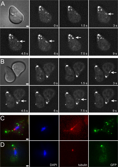
Figure 4.
Fus2p movement and localization are independent of microtubules. MY9184 expressing Fus2-GFP expressed from its own promoter was treated with α-factor for 1.5 h and either mock treated (A and C) or treated with nocodazole (B and D) for 10 min. Live cell image stacks were acquired (A and B). Arrows indicate directed movement of Fus2p-GFP puncta. Alternatively, cells were fixed and stained with anti-tubulin antibody (C and D) to demonstrate microtubule depolymerization. Bars, 1 μm.

