Figure 6.
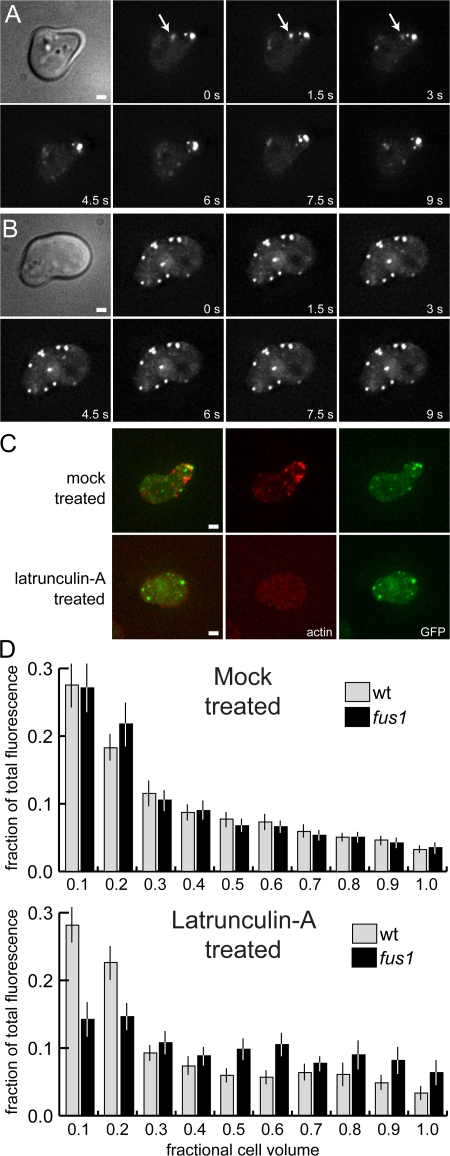
Cortical localization is dependent on both actin and Fus1p. (A and B) Fus2p-GFP localization is disrupted by lat-A in a fus1 mutant. MY9217 was treated with α-factor for 1.5 h and either mock treated or treated with lat-A as in Fig. 5. (A) The fus1 mutation had no effect on the transport or localization of Fus2p-GFP. Arrows indicate rapid movement of Fus2p-GFP dots. (B) Treatment with lat-A (10 min) leads to a rapid loss of Fus2p-GFP localization at the shmoo tip. (C) Texas red–phalloidin staining of fixed cells after lat-A treatment demonstrating actin depolymerization. Bars, 1 μm. (D) Distribution of GFP fluorescence in wild-type and fus1 mutant shmoos. The fraction of total fluorescence was measured for every 1/10 of the cell, starting at the shmoo tip. 50 cells were measured for each strain and condition. Bars represent the mean values; error bars indicate 95% confidence intervals.

