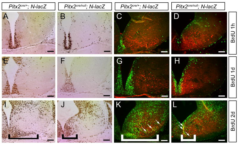
Figure 5.
Hypothalamic Pitx2cre/null; N-lacZ neurons fail to migrate. E14.5 Pitx2cre/+; N-lacZ (A, C, E, G, I, K) and Pitx2cre/null; N-lacZ (B, D, F, H, J, L) embryos were exposed to BrdU by intraperitoneal injection 1 hour (A–D), 1 day (E–H), or 2 days (I–L) prior to collection, and transverse sections processed for anti-BrdU immunohistochemistry (A, B, E, F, I, J) or double-label immunofluorescence with anti-BrdU and anti-β-galactosidase (C, D, G, H, K, L). There were no differences in BrdU labeling between Pitx2cre/+ and Pitx2cre/null mutants injected with BrdU 1 hour (A–D) or 1 day (E–H) prior to analysis. Pitx2cre/null mutants injected with BrdU 2 days prior to analysis (I–L) exhibited fewer BrdU positive cells in the lateral hypothalamus (subthalamic nucleus region) compared with Pitx2cre/+ embryos (bracketed areas). There was no cellular colocalization between BrdU and β-gal in embryos injected at E14.5 (C, D) or E13.5 (G, H), whereas double-labeled cells were present in embryos injected at E12.5 (white arrows in K and L). Scale bars = 100 μm.

