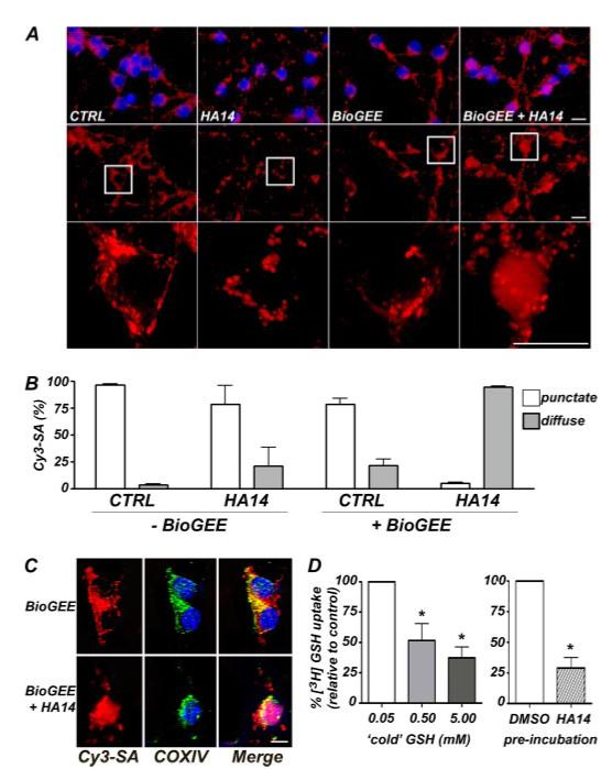
FIGURE 3. The BH3 mimetic, HA14-1, displaces the mitochondrial GSH pool in CGNs and inhibits GSH transport into isolated mitochondria.

A, the cellular distribution of a BioGEE probe was visualized with streptavidin conjugated to Cy3 (red). In CGNs that were not loaded with BioGEE (left two columns), Cy3-streptavidin (Cy3-SA) bound to biotinylated proteins in the mitochondria, and exposure to HA14 had no effect on the localization of staining. However, in neurons that were loaded with BioGEE for 1 h prior to treatment (right two columns), the Cy3-SA staining showed a redistribution from a punctate and primarily mitochondrial localization in CTRL cells, to a much more diffuse, cytoplasmic staining pattern in HA14-treated cells. The nuclei were stained with DAPI (blue). Top row, Cy3-SA and DAPI; middle row, Cy3-SA alone; bottom row, the areas demarcated by the boxes were magnified approximately four times. Scale bars, 10 microns. B, quantitation of punctate versus diffuse Cy3-SA staining (as described in A) in CGNs that were either unloaded or loaded with BioGEE for 1 h prior to HA14 (15 μm) treatment for 2 h. The data shown are the means ± S.E. for four separate experiments in which ∼250 cells were quantified per experiment. C, BioGEE, labeled by Cy3-SA staining (red), co-localized with the integral mitochondrial membrane protein, COX IV (green), in CTRL CGNs but not in cells treated with the Bcl-2 inhibitor, HA14. The nuclei were stained with DAPI (blue). Scale bar,10 microns. D, mitochondria isolated from whole neonatal rat brain were incubated with 0.5 μCi of [3H]GSH for 15 s at room temperature. Increasing the concentration of unlabeled (cold) reduced GSH in the incubation buffer competed with the [3H]GSH and caused a decrease in its uptake into mitochondria (left bar graph). *, p < 0.01 versus the lowest concentration of cold GSH (0.05 mm), which was arbitrarily set at 100% [3H]GSH uptake. Preincubation for ∼20 min with the BH3 mimetic HA14 (20 μm) significantly blocked [3H]GSH uptake into isolated mitochondria when assayed in the presence of 0.05 mm cold GSH (right bar graph). *, p < 0.01 versus Me2SO vehicle preincubation. The results shown are the means ± S.E. of three experiments, each performed in triplicate. CTRL, control.
