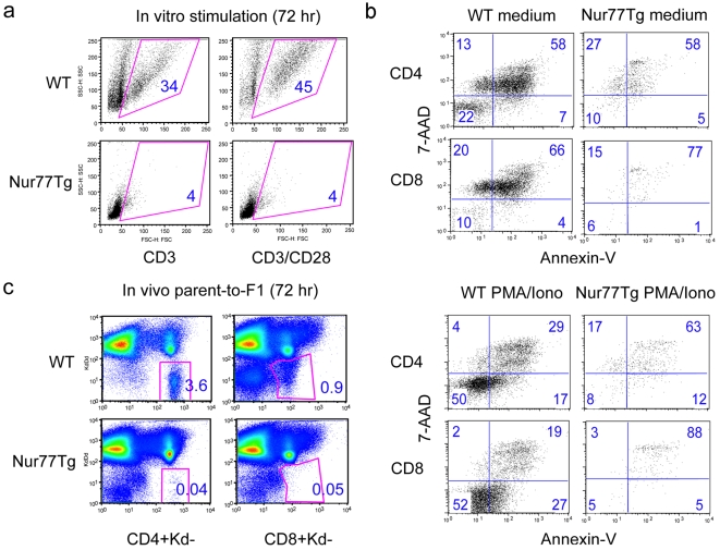
Figure 2. Increased apoptosis of Nur77Tg T cells upon activation in vitro and in vivo.
Splenocytes from WT or Nur77Tg mice were stimulated with PMA/ionomycin for 72 h, followed by flow cytometry using T cell markers and Annexin-V and 7-AAD staining. (a) Live cells were gated based on forward vs. side scatter, and (b) T cell apoptosis was based on Annexin-V and 7-AAD staining. (c) CFSE-labeled B6 or Nur77Tg spleen and lymph node cells were adoptively transferred into B6/DBA F1 mice. After 72 h, donor-derived live CD4+ or CD8+ T cells were identified by gating on H-2Kd (-) and H-2Dd (-) cells; figure in each square indicates percentage of the gated population. Data are representative of 3 experiments with similar results.

