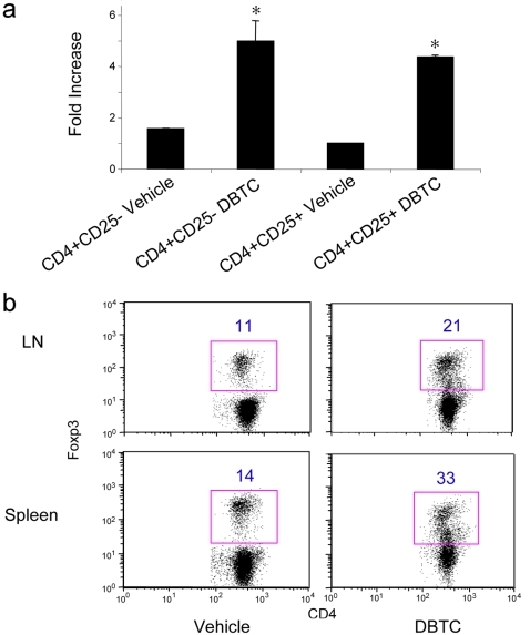
Figure 7. DBTC induces Nur77 and differentially promotes non-Treg vs. Treg cell apoptosis.
(a) WT CD4+CD25− and CD4+CD25+ T cell populations were fractionated using magnetic beads (>90% purity) and cultured for 4 h in RPMI medium plus 3 µM DBTC or ethanol alone (final concentration of 0.1% ethanol). Nur77 mRNA expression was detected by qPCR; data are expressed as fold increase (mean±SD) above control set as 1. DBTC treatment increased Nur77 mRNA in both cell populations (p<0.05 for CD4+CD25− vs. control, p<0.01 for CD4+CD25 vs. control), but levels of induction at 4 h were not significantly different (p>0.05) for the groups treated with DBTC. (b) C57BL/6 mice were fed DBTC (60 mg/kg/day) for 3 d, sacrificed and single cell suspensions from spleens and lymph nodes were stained with T cell markers and Foxp3 mAb. Flow cytometric analysis of Foxp3 expression in the CD4+ T cell population is representative of 4 mice/group, and the percentage of the gated population is indicated.

