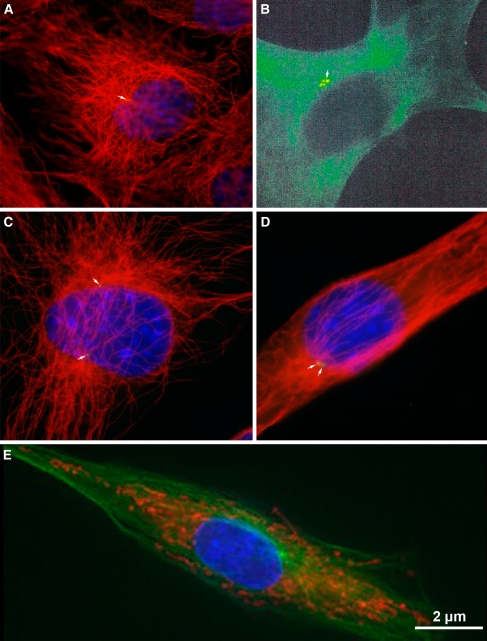
Fig. 4.
In interphase, a single centrosome is juxta-positioned to the nucleus. a, c, and d show small GFP-centrin-labeled centrosomes in mouse 3T3 cells. Microtubules are detected with α-tubulin and shown in red; b is of an LNCaP prostate cancer cell labeled with human autoimmune antibody SPJ displaying multiple centrosomal foci perhaps indicating centrosome abnormalities; e shows a porcine fibroblast cell labeled with γ-tubulin to detect the centrosome and Mitotracker Rosamine to detect mitochondria. Microtubules are shown in green; b reprinted with permission from Schatten et al. 2000b

