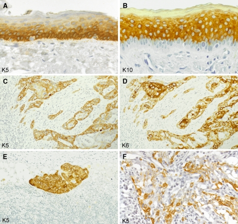
Fig. 4.
Keratins in stratified squamous epithelia and squamous cell carcinomas (paraffin sections of human tissues; avidin–biotin complex peroxidase staining). In the epidermis as an example of a normal stratified squamous epithelium, the basal cell layer contains abundant keratin K5 (a) whereas the differentiating suprabasal compartment strongly stains for K10 (b; note the negative basal cell layer). Lymph node metastasis of a squamous cell carcinoma of the head and neck region, expressing K5 (c; more intensely in the peripheral tumor cell layers) as well as K6 (d; particularly strongly in central tumor cells) as signs of their keratinocyte origin. Keratin K5 is also maintained in a lymph node micrometastasis of a squamous cell carcinoma of the head and neck region (e) and in a lymph node metastasis of an undifferentiated nasopharyngeal carcinoma with dissociated growth pattern of the tumor cells (f), in these examples being a diagnostically helpful feature. Magnifications: a, b ×160; c–e ×80; f ×140

