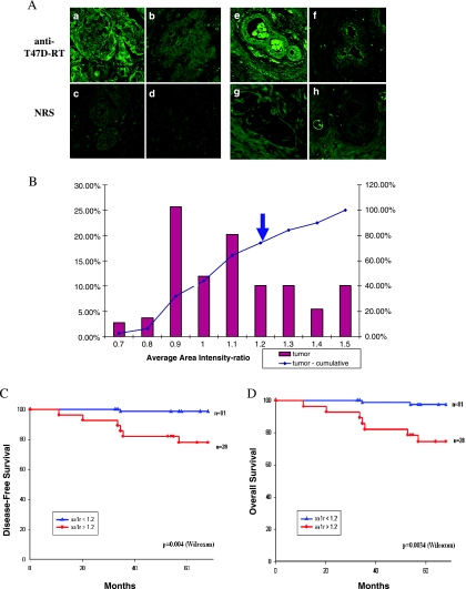
Figure 5.
HERV-K T47D-RT expression in human breast carcinoma tissues. (A) Paraffin-embedded sections from breast carcinoma biopsies were subjected to indirect immunoflourescence staining using anti-T47D-RT antibody (a, b, e, and f) or NRS (normal rabbit serum) (c, d, g, and h) and analyzed by CLSM. Representative image pairs of anti-T47D-RT and NRS are presented: RT-positive (a, c) or RT-negative (b, d) tumor tissue; RT-positive (e, g) or RT-negative (f, h) normal adjacent tissue serial sections. (B) The distribution of the tumors' HERV-K T47D-RT staining level (AAI ratio; bars) and their cumulative percentages (lines). The cutoff point between positive and negative staining is marked by arrow (AAI = 1.2). (C) Kaplan-Meier analysis of disease-free survival for two groups of patients: HERV-K T47D-RT-positive (as measured by AAI ratios), at levels above the cutoff of 1.2 (red line); and HERV-K T47D-RT-negative, at levels below the cutoff (blue line). (D) Kaplan-Meier analysis of overall survival for the same two groups of patients as in panel (C): HERV-K T47D-RT-positive (red line) and HERV-K T47D-RT-negative (blue line). P values were calculated using log-rank and Wilcoxon tests.

